Loading...
Blood & the Circulatory System
Quiz by Devon Roesener
Tag the questions with any skills you have. Your dashboard will track each student's mastery of each skill.
Carry oxygen and nutrients throughout the body
An enlargement of the spleen
What are the three arteries that supply blood to the spleen

Person trained to draw blood from a patient.
Red blood cells appear faded (lose their color)
An inherited disorder that interferes with the blood’s ability to carry oxygen.
If both parents are carriers for Cooley's anemia, what are the chances their children will have Cooley's anemia?
Blood is what type of tissue?
How many main components are there to blood?
What are the two main components of blood?
The shape of normal blood cells.
Red blood cells
White blood cells
Platelets
Made from a protein called hemoglobin (Hb) which contains iron used to transport oxygen.
Normal red blood cells do not contain a(n) . . .
-formation of blood cells
- occurs in the bone marrow
- an error in the genetic code can cause the protein to be abnormally shaped
A hormone that increases production of RBC’s.
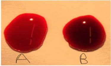
Which is the deoxygenated blood spot?
Immune function (several types)
Attacks bacteria through phagocytosis
Attacks parasites and can be present in response to allergies
Active in injury sites. Releases heparin to prevent clotting & histamines to increase inflammation
Initiates immune response to viruses
Involved in attacking viruses & abnormal tissues
Initiates clotting process
In 1667, this physician was the first to complete an animal to human blood transfusion.
1901 - Austrian _____ discovered human blood groups, which we now call blood types.
Blood cells have markers on their surface called
In addition to the ABO groups, blood type can have another protein on the surface. If blood cells have this factor, they are described as positive. Cells without this surface protein are described as negative.
Antigens generate antibodies (antigen is short for ___-___).
___ are proteins that circulate in the blood and can attach to foreign invaders in the blood.
When antibodies attach to blood cells, the blood cells clump together, this is known as ___.
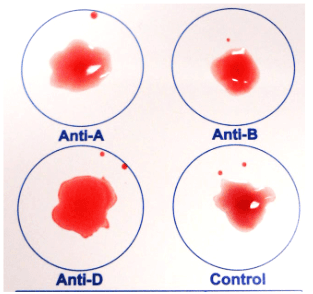
What blood type is represented by the picture?
Why is type O blood considered the universal donor?
Antigens produced by the fetus can generate antibodies in the mother.
What is the rarest blood type?
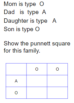
What are the possible genotypes for type A blood?
What percentage of their children will have Type A blood?
The liquid portion of blood is _____ water.
Plasma proteins - albumins
plasma proteins - globulins
plasma proteins - fibrinogen
Involves the coagulation and clotting of the blood to seal the site of damage.
Key events in hemostasis.
An enzyme in blood plasma that causes the clotting of blood by converting fibrinogen to fibrin.
The thickening of blood to form a clot (hematoma).
blood clot (abnormal)
When the clot moves to another place.
When a blood clot goes to the lungs.
Bleeder's disease
The average adult has how many pints of blood in their body.
The ratio of the volume of red blood cells to the total volume of blood.
A type of blood cancer.
___ = a cancer that progresses quickly
___ refers to its effect on cells that turn into healthy white blood cells
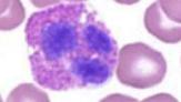
White blood cells that have a granular cytoplasm.
White blood cells that lack a granular cytoplasm.
Granulocytes
Agranulocytes
White bloods cells are ___ than red blood cells.
60% of WBC.
30% of WBC
Basophil
What type of WBC is depicted in the picture?
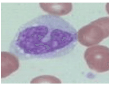
What type of WBC is depicted in the picture?
What type of WBC is depicted in the picture?
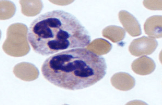
What type of WBC is depicted in the picture?

Delivers blood to all body cells and carries away waste.
Eliminates carbon dioxide and oxygenates blood (lung pathway).
Located in the ___, between the 2nd rib and the 5th intercostal space.
The distal end of the heart is called . . .
#11

circumflex coronary artery
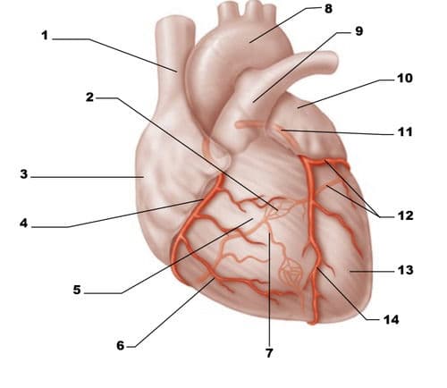
#4

left anterior descending coronary artery

Encloses the heart (like a bag) and has 2 layers.
Inner layer of the fibrous pericardium.
Outer layer of the fibrous pericardium.
Contains fluid reducing friction around the heart.
Outer layer of the heart wall.
Middle layer of the heart wall.
Thin inner lining, within chambers of the heart.
Thin upper chambers that receive blood returning to the heart through veins.
Thick, muscular lower chambers. Pumps blood out of the heart through arteries (away).
Separates the right and left sides of the heart.
The bicuspid or mitral valve.
The tricuspid valve.
Valve between the left ventricle and aorta.
Valve between the right ventricle and pulmonary artery.
Pulmonary Semilunar Valve

#1

#3
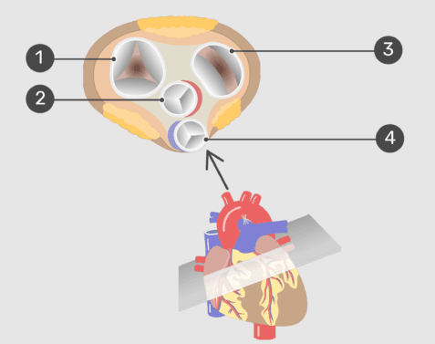
Aortic Semilunar Valve

The cusps (flaps) of the bicuspid and tricuspid valves are anchored to the ventricle walls by fibrous “cords” called . . .
#9

#2

#7

#1

#8

#3
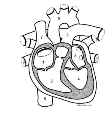
#6

#4

#5

One complete heartbeat.
Contraction of the heart chamber.
Relaxation of a heart chamber.
Blood pressure cuffs measure the ___ in the vessels.
During systole, this force is the greatest, on a blood pressure reading, this is the ___ .
Diastole is the smaller number, when the ___ .
Average human blood pressure.
Instrument to listen and measure heart sounds.
Places where arteries come close to your skin--on your body.
Pulse point located on your neck.
Pulse point located on your wrist.
Pulse point located on your arm.
A recording of the electrical events (changes) during a cardiac cycle.
Depolarization of the atria (atrial contraction – systole)
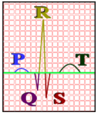
Depolarization of the ventricles (ventricular contraction, systole)

Repolarization of the ventricles

Fast heart rate
slow heart rate
irregular heart rate
Heart rate is controlled by the ___ within the medulla oblongata.
Strong elastic vessels which carry blood moving away from the heart. Smallest ones are arterioles which connect to capillaries.
Thinner, less muscular vessels carrying blood toward the heart.
Smallest veins are called ___ which connect to capillaries and contain valves.
Allows exchange of materials (oxygen, nutrients) between blood and tissues.
Circular, valve-like muscle at arteriole-capillary junction.
Narrowing of the vessel.
Expanding of the vessel.
Largest artery.
Splits into left and right, both lead to the lungs.
Returns blood from the lungs to the heart.
Returns blood from the head and body to the heart.
Supply's blood to the heart itself.
branches into the:
Right Subclavian ( supplies blood to the arms)
Left Subclavian Artery – supplies blood to the left arms
Right Common Carotid (bicarotid)
The device shocks the heart and allows it to re-establish its normal; can also be used to start a heart that has stopped.
CPR
Unusual sound heard during a heartbeat Sometimes they sound like a whooshing or swishing noise.
An obstruction to the coronary artery, commonly called a “heart attack”
High blood pressure, the force within the arteries is too high.
Is a localized, blood-filled balloon-like bulge in the wall of a blood vessel.
Blood flow to the brain is cut off.
Valve or aorta is narrowed, limiting blood flow.
A hole exists between the two sides of the heart (septum).
Deposits of fatty materials such as cholesterol form a “plaque” in the arteries which reduces blood flow.