Loading...
Chapter 12 - Part III - Principles of Anatomy and Physiology
Quiz by Andre McBean
Tag the questions with any skills you have. Your dashboard will track each student's mastery of each skill.
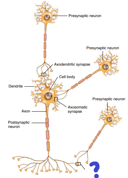
A ____________ is a region where communication occurs between two neurons or between a neuron and an effector cell (muscle cell or glandular cell)
The nerve cell that is in the position of sending the signal from its axon hillock before the synapse is called the ______________ neuron.
The nerve cell that is in the position of receiving the signal after the synapse is called the ______________ neuron.
Where the axon meets the dendrites at the synapse is called _________.
Where the axon meets the cell body at the synapse is called _________.
Take a look at the image and all the synaptic connections. Where the axon meets axon at the synapse is called _________.
There are two types of synapses based on the signal being sent: electrical synapse and chemical synapse.
Watch the following video: youtu.be/vpjp-2IJ_fo and youtu.be/TQ6n3rZ_rL8 and youtu.be/WhowH0kb7n0
At an electrical synapse, action potentials conduct directly between the plasma membranes of adjacent neurons through structures called gap junctions. Each gap junction contains a hundred or so tubular connexons, which act like tunnels to connect the cytosol of the two cells directly. Gap junctions are common in visceral smooth muscle, cardiac muscle, and the developing embryo. They also occur in the brain.
Because action potentials conduct directly through gap junctions, electrical synapses are slower than chemical synapses.
Electrical synapses are slower at synchronization. In other words, they take longer to find the correct path of communication, or coordination.
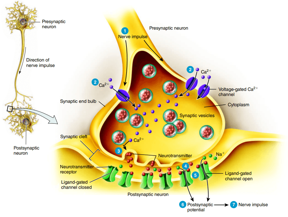
Although the plasma membranes of presynaptic and postsynaptic neurons in a chemical synapse are close, they do not touch. They are separated by the ______________, a space of 20–50 nm* that is filled with interstitial fluid.
Nerve impulses cannot conduct across the synaptic cleft, so an alternative, indirect form of communication occurs. In response to a nerve impulse, the presynaptic neuron releases a ___________________ that diffuses through the fluid in the synaptic cleft and binds to receptors in the plasma membrane of the postsynaptic neuron.
The postsynaptic neuron receives the chemical signal and in turn produces a _________________potential, a type of graded potential.
The presynaptic neuron converts an electrical signal (nerve impulse) into a chemical signal (released neurotransmitter). The postsynaptic neuron receives the chemical signal and in turn generates an electrical signal (postsynaptic potential).
The time required for these processes at a chemical synapse, a ______________ of about 0.5 msec, is the reason that chemical synapses relay signals more slowly than electrical synapses.
When a depolarizing (increasing value to positive >-55mV) postsynaptic potential (electrical signal at the postsynaptic membrane) reaches threshold, it triggers a nerve impulse in the axon of the postsynaptic neuron. Of the seven steps of chemical signaling, which step is this?
Complete the following quiz: https://www.purposegames.com/game/presynaptic-neuron-to-post-synaptic-neuron
At most chemical synapses, only one-way information transfer can occur—from a presynaptic neuron to a postsynaptic neuron or an effector, such as a muscle fiber or a gland cell. Which of the following is responsible for the one way signaling?
A neurotransmitter can only cause an excitatory (stimulating to threshold) graded potential.
A depolarizing postsynaptic potential is called an _____________ postsynaptic potential.
A hyperpolarizing postsynaptic potential is called an _____________ postsynaptic potential.
IPSP and EPSP are both different electrical impulses that are generated as a signaling response.
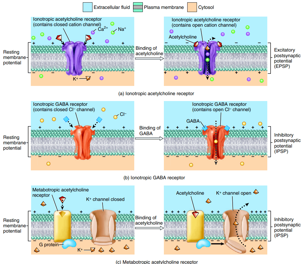
When a neurotransmitter binds to the protein receptor (neurotransmitter receptor), either an excited, or inhibited graded potential occurs. An excited potential (on) is called an EPSP (excitatory postsynaptic potential), and an inhibited potential (off) is called and IPSP (inhibitory postsynaptic potential). However, there are more than one type of neurotransmitter receptors. A _______________ receptor is a type of neurotransmitter receptor that contains a neurotransmitter binding site and an ion channel, both as a component of the same protein unit.
What type of channel is the ionotropic receptor?
Many excitatory neurotransmitters bind to ionotropic receptors that contain ____________ channels. EPSPs result from opening these ___________ channels. When __________channels open, they allow passage of the three most plentiful cations (Na+, K+, and Ca2+) through the postsynaptic cell membrane, but Na+ inflow is greater than either Ca2+ inflow or K+ outflow, and the inside of the postsynaptic cell becomes less negative (depolarized).
Many inhibitory neurotransmitters bind to ionotropic receptors that contain ___________ channels (Figure 12.24b). IPSPs result from opening these _______ channels. When ________ channels open, a larger number of chloride ions diffuse inward. The inward flow of Cl− ions causes the inside of the postsynaptic cell to become more negative (hyperpolarized).
Match the following channels to their effect on the postsynaptic membrane:
A ______________ receptor is a type of neurotransmitter receptor that contains a neurotransmitter binding site but lacks an ion channel as part of its structure. However, it's not that there isn't the presence of any ion channels. It's that they are not bound as one structure quite like ionotropic receptor proteins being one structure. A ____________ receptor is instead coupled (linked) to a separate ion channel by a type of membrane protein.
The protein that links a metabotropic receptor to the ion channel is called a _______ protein. This protein is responsible for signaling the ion channel to act when the ________ protein detects the activity of the metabotropic receptor being bonded to.
Metabotropic receptors mostly respond by opening K+ ion channels to release potassium ions thus hyperpolarizing the cytoplasm. Therefore, which type of neurotransmitter signal does it mostly respond to?
The same neurotransmitter can be excitatory at some synapses and inhibitory at other synapses, depending on the structure of the neurotransmitter receptor to which it binds.
Removal of the neurotransmitter from the synaptic cleft is not necessary for normal synaptic function. As there is a built-in tolerance for low levels of neurotransmitter presence.
Since it is important that neurotransmitters are removed for proper function, match the definitions of each type of removal method with the terms:
A reminder that summation is the electrical excitement of a neuron multiple times in a row. It can be by just one neuron to the next neuron, or it can be multiple neurons "zapping" just one other neuron. That's when a summation is formed -- a graph of the multiple "shocks" the neuron received.
A typical neuron in the CNS receives input from 1000 to 10,000 synapses. Integration of these inputs involves summation of the postsynaptic potentials that form in the postsynaptic neuron. Recall that summation is the process by which graded potentials add together. The greater the summation of EPSPs, the greater the chance that threshold will be reached. At threshold, one or more action potentials arise. There are two types of summation: __________summation and ____________summation. Use "and" to combine both answers. Don't use the word summation.
________________ summation is summation of postsynaptic potentials in response to stimuli that occur at different locations in the membrane of a postsynaptic cell at the same time.
________________ summation is summation of postsynaptic potentials in response to stimuli that occur at the same location in the membrane of a postsynaptic cell but at different times.
Group the following based on fit:
Review the following image to proceed and answer the next question:
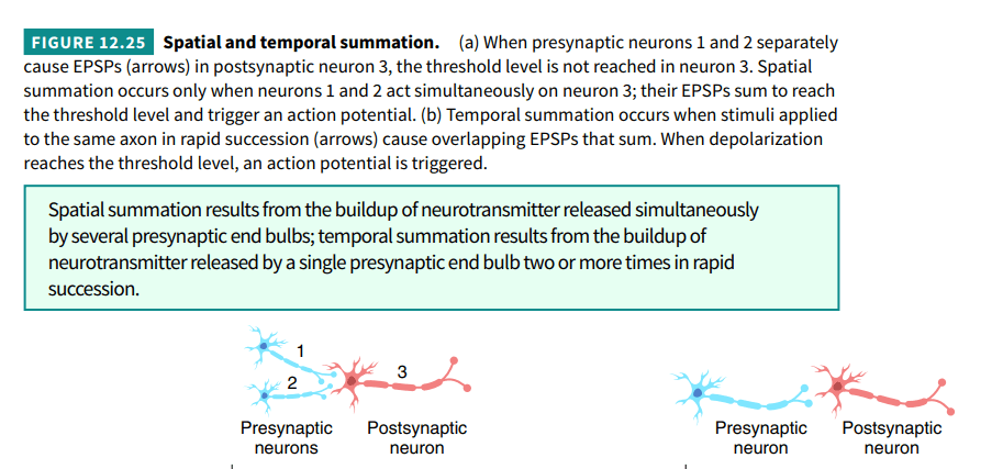
Spatial summation is the result of many presynaptic signals generated at the same time. Whereas, temporal is from one presynaptic signal, hence why they are distinct, one at a time on a graph of the electrical impulses.
EPSP stand for ____________________________.
IPSP stand for ____________________________.
If the total excitatory effects are greater than the total inhibitory effects but less than the threshold level of stimulation, will the EPSP reach the threshold?
If the total excitatory effects are greater than the total inhibitory effects, a threshold is reached. What is the name of the response generated by the output neuron (postsynaptic)?
If the total inhibitory effects are greater than the excitatory effects, the membrane depolarizes.
There are neurons that can secrete hormones called neurosecretory hormones in the brain.
How many classes of neurotransmitters are there?
Group the following into the two classes of neurotransmitters: small-molecule neurotransmitters and neuropeptides.
The enzyme ______________________ inactivates ACh by splitting it into acetate and choline fragments.
Most excitatory neurons in the CNS and perhaps half of the synapses in the brain communicate via glutamate.
Gamma-aminobutyric acid (GABA) and glycine are important inhibitory neurotransmitters. The binding of GABA to ionotropic receptors opens Cl− channels. GABA is found only in the CNS, where it is the most common inhibitory neurotransmitter. As many as one-third of all brain synapses use GABA. Antianxiety drugs such as diazepam (Valium®) enhance the action of GABA. Like GABA, the binding of glycine to ionotropic receptors opens Cl− channels. About half of the inhibitory synapses in the spinal cord use the amino acid glycine, the rest use GABA.

Match the following neurotransmitter/biogenic amines with their descriptions:
Inactivation of catecholamines occurs via reuptake into synaptic end bulbs. Then they are either recycled back into the synaptic vesicles or destroyed by enzymes. The two enzymes that break down catecholamines are catechol-O-methyltransferase (COMT), and monoamine oxidase (MAO).
Neurotransmitters consisting of 3 to 40 amino acids linked by peptide bonds called ___________________. They are formed in the neuron cell body, packaged into vesicles, and transported to axon terminals.
Scientists discovered that certain brain neurons have plasma membrane receptors for opiate drugs such as morphine and heroin due to the drug effect on brain activity. This sent them on a hunt to discover the receptors and study them. As a result, they discovered the very first neuropeptide, enkephalins. They are so potent that they are about 200 times stronger then even morphine. Scientists continued on the hunt for other opioid peptides which lead them to endorphins and dynorphins. Scientists ultimately concluded that these receptors are responsible for satisfying the body's needs for painkiller response. These neuropeptides have also been linked to improved memory and learning; feelings of pleasure or euphoria; control of body temperature; regulation of hormones that affect the onset of puberty, sexual drive, and reproduction. On the other hand, the neuropeptide _____________ is released in response to pain receptors.
Classifying neural circuits based on the arrangement of neurons and their firing patterns. Identify the type of circuit in A:
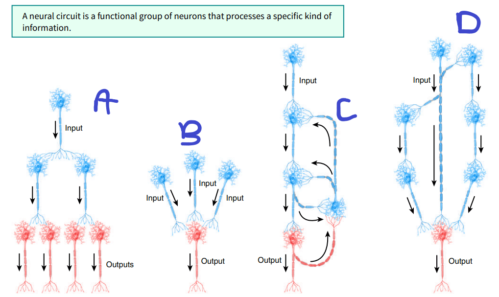
Classifying neural circuits based on the arrangement of neurons and their firing patterns. Identify the type of circuit in D:
Classifying neural circuits based on the arrangement of neurons and their firing patterns. Identify the type of circuit in C:
Classifying neural circuits based on the arrangement of neurons and their firing patterns. Identify the type of circuit in B:
Match the neural circuit with the likely activity:
The neuron's arrangement capability to change readily based on experience. At the level of individual neurons, the changes that can occur include the sprouting of new dendrites, synthesis of new proteins, and changes in synaptic contacts with other neurons.
Despite neuron plasticity, mammalian neurons have very limited powers of regeneration, the capability to replicate or repair themselves.
In the CNS, damage to dendrites and myelinated axons may be repaired if the cell body remains intact and if the Schwann cells that produce myelination remain active. In the PNS, little or no repair of damage to neurons occurs.
Even when the cell body remains intact, a severed axon cannot be repaired or regrown.
__________________ is the birth of new neurons from undifferentiated stem cells.
Until recently, the dogma (belief) in humans and other primates was “no new neurons” in the adult brain. Then, in 1992, researchers published their unexpected finding that the hormone-like protein _______________________ stimulated cells taken from the brains of adult mice to proliferate into both neurons and astrocytes. Previously, __________________was known to trigger mitosis in a variety of nonneuronal cells and to promote wound healing and tissue regeneration. In 1998, scientists discovered that significant numbers of new neurons do arise in the adult human hippocampus, an area of the brain that is crucial for learning.
The nearly complete lack of neurogenesis in other regions of the brain and spinal cord seems to result from two factors: (1) inhibitory influences from neuroglia, particularly oligodendrocytes, and (2) absence of growth-stimulating cues that were present during fetal development. Axons in the ___________ are myelinated by oligodendrocytes rather than Schwann cells, and this ____________myelin is one of the factors inhibiting regeneration of neurons. Perhaps this same mechanism stops axonal growth once a target region has been reached during development. Also, after axonal damage, nearby astrocytes proliferate rapidly, forming a type of scar tissue that acts as a physical barrier to regeneration. Thus, injury of the brain or spinal cord usually is permanent.
In the peripheral nervous system (PNS), axons and dendrites that are associated with a neurolemma may repair themselves only under certain circumstances. Which of the following are part of the limited criteria for repair in the PNS to happen:
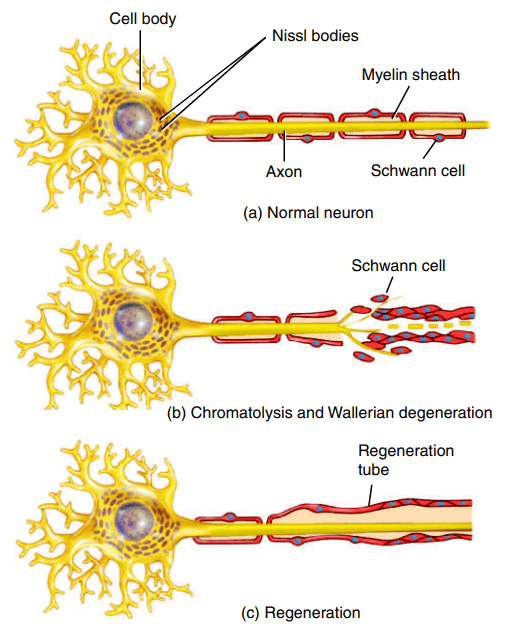
When there is damage to an axon, changes usually occur both in the cell body of the affected neuron and in the portion of the axon distal to the site of injury. Changes also may occur in the portion of the axon proximal to the site of injury. About 24 to 48 hours after injury to a process of a normal peripheral neuron, the Nissl bodies break up into fine granular masses. This alteration is called _____________________.
Degeneration (breakdown/destruction) of the distal portion of the axon and myelin sheath is called ______________________.
New axons cannot grow if the gap at the site of injury is too large or if the gap becomes filled with collagen fibers. During the first few days following damage, buds of regenerating axons begin to invade the tube formed by the Schwann cells. Axons from the proximal area grow at a rate of about 1.5 mm (0.06 in.) per day across the area of damage, find their way into the distal regeneration tubes, and grow toward the distally located receptors and effectors. Thus, some sensory and motor connections are reestablished, and some functions are restored. In time, the Schwann cells form a new myelin sheath.
Watch the following video: youtu.be/4009Nv4C4NY
Match each of the neurodegenerative conditions with their descriptions:
