Loading...
Chapter 4 - Animal - Listening Exercise (3A)
Quiz by Jessica Shandy
Customize this quiz to suit your class
Instantly translate to 100+ languages
Tag the questions with any skills you have. Your dashboard will track each student's mastery of each skill.
Give this quiz to my class
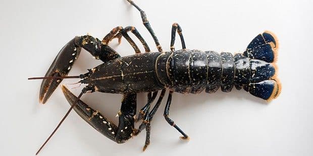
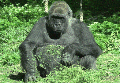
Choose the correct picture according to the recording.
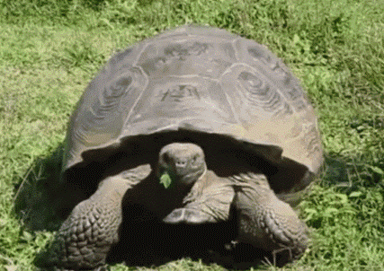
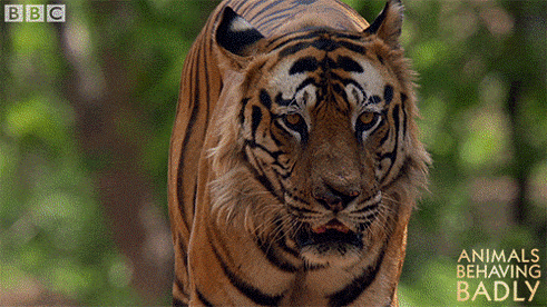
Choose the correct picture according to the recording.
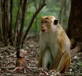
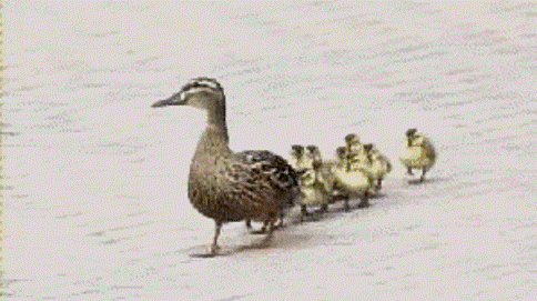
Choose the correct picture according to the recording.
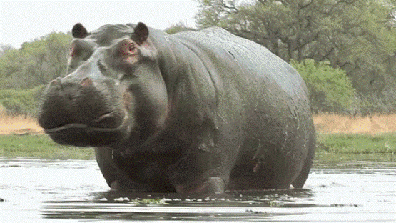
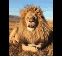
Choose the correct picture according to the recording.
Choose the correct picture according to the recording.
Choose the correct picture according to the recording.
Choose the correct picture according to the recording.
Choose the correct picture according to the recording.
Choose the correct picture according to the recording.
Choose the correct picture according to the recording.
Choose the correct picture according to the recording.
Choose the correct picture according to the recording.
Choose the correct picture according to the recording.
Choose the correct picture according to the recording.
Choose the correct picture according to the recording.
Choose the correct picture according to the recording.
Choose the correct picture according to the recording.
Choose the correct picture according to the recording.
Choose the correct picture according to the recording.
According to the audio, choose the correct sentence!
According to the audio, choose the correct sentence!
According to the audio, choose the correct sentence!