Loading...
The structure and function of the skin
Quiz by Maria Owen
Customize this quiz to suit your class
Instantly translate to 100+ languages
Tag the questions with any skills you have. Your dashboard will track each student's mastery of each skill.
Give this quiz to my class
Which of the following is the outside of the skin
Subcutaneous fat layer
Epidermis
Dermis
Nerve endings
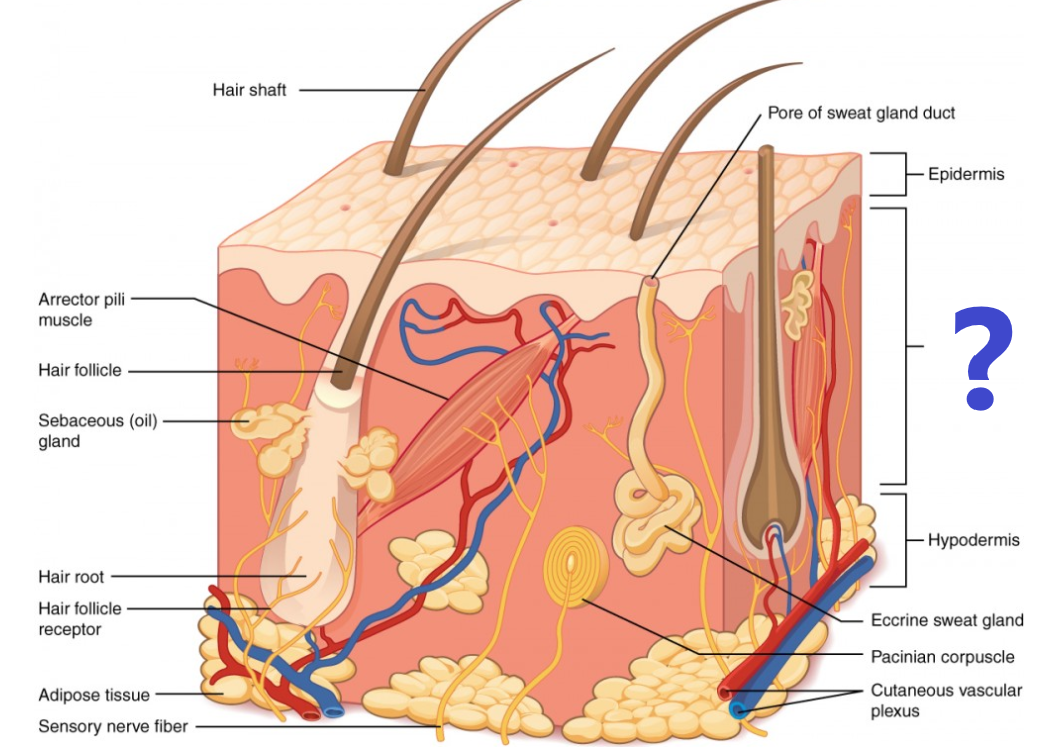
Which layer of the skin has not been named on the diagram has not been named? (blue question mark)
Dermis
Epidermis
Pores
Subcutaneous fat
Which of the following is the outside of the skin
Which layer of the skin has not been named on the diagram has not been named? (blue question mark)

The _______________ is the layer of skin that contains nerve endings, blood vessels, oil glands, and sweat glands.
_____________ are tiny holes in the skin that allow sweat to escape.
Hair follicles have their roots in which layer of skin?
The skin is the largest organ of the human body.
Which of the following is not a job of the epidermis?
The skin is involved in producing which of the following vitamins?
Which layer contains the nerves & blood vessels?
Which layer of the skin does NOT contain blood vessels?