
Ácidos Nucleicos TEST
Quiz by Pilar Iniesto López
Customize this quiz to suit your class
Instantly translate to 100+ languages
Tag the questions with any skills you have. Your dashboard will track each student's mastery of each skill.
Give this quiz to my class
Las bases púricas son ....
la citosina y la timina
la adenina y la timina
la citosina y la guanina
la adenina y la guanina
Un nucleótido es
cada una de las bases de los ácidos nucleicos
el monómero que forma las proteínas
la unión del monosacárido y la base nitrogenada
el monómero que forma los ácidos nucleicos
Las bases púricas son ....
Un nucleótido es
Los nucleótidos se unen entre sí para formar un polinucleótido mediante enlaces...
El análisis de 4 fragmentos de una molécula de ADN extraída de una bacteria ha proporcionado las composiciones de bases a, b, c y d ¿Cuál desnaturalizará a mayor temperatura?
Los ácidos nucleicos son...
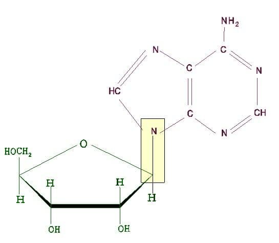
¿A qué OH del azúcar, de los que se observan en la figura, se une el OH del el ácido fosfórico para formar un nucleótido?

En el ADN bicatenario se cumple la siguiente norma
El enlace indicado con un recuadro en la figura es un enlace...

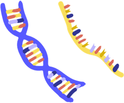
La desoxirribosa se diferencia de la ribosa en que...
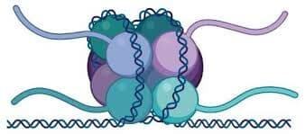
¿Cómo se llama en general lo que se observa en la figura si representa parte de la estructura terciaria de un ácido nucleico?

LAB_TEST FOR NUCLEIC ACIDS (FINALS)
Lipids are large, nonpolar organic molecules. They do not dissolve in water. Lipids include triglycerides (trie-GLIS-uhr-IEDZ), phospho- lipids, steroids, waxes, and pigments. Lipid molecules have a higher ratio of carbon and hydrogen atoms to oxygen atoms than carbohydrates have. Because lipid molecules have larger numbers of carbon-hydrogen bonds per gram than other organic com- pounds do, they store more energy per gram. Fatty Acids Fatty acids are unbranched carbon chains that make up most lipids. Figure 3-10 shows that a fatty acid contains a long carbon chain (from 12 to 28 carbons) with a carboxyl group, —COOH, attached at one end. The two ends of the fatty-acid molecule have different properties. The carboxyl end is polar and is thus hydrophilic or attracted to water molecules. In contrast, the hydro- carbon end of the fatty-acid molecule is nonpolar. This end tends not to interact with water molecules and is said to be hydrophobic (HIE-droh-FOH-bik), or “water fearing.” In saturated fatty acids, such as palmitic acid, which is shown in Figure 3-10, each carbon atom is covalently bonded to four atoms. The carbon atoms are in effect full, or saturated. In contrast, linoleic acid, also shown in Figure 3-10, has carbon atoms that are not bonded to the maximum number of atoms to which they can bond. Instead, they have formed double bonds within the carbon chain. This type of fatty acid is said to be unsaturated. Triglycerides Three classes of lipids important to living things contain fatty acids: triglycerides (fats), phospholipids, and waxes. A triglyceride is composed of three molecules of fatty acid joined to one molecule of the alcohol glycerol. Saturated triglycerides are composed of saturated fatty acids. They typically have high melting points and tend to be hard at room temperature. Common dietary saturated triglycerides include butter and fats in red meat. In contrast, unsaturated triglycerides are composed of unsaturated fatty acids and are usually soft or liquid at room temperature. Unsaturated triglycerides are found primarily in plant seeds where they serve as an energy and carbon source for germinating plants. Phospholipids Phospholipids have two, rather than three, fatty acids attached to a molecule of glycerol. They have a phosphate group attached to the third carbon of the glycerol. As shown in Figure 3-11, the cell membrane is made of two layers of phospholipids, called the lipid bilayer. The inability of lipids to dissolve in water allows the mem- brane to form a barrier between the inside and outside of the cell. Hydrophilic “head” Phospholipids Hydrophobic “tail” Phospholipids Water Water The lipid bilayer of a cell membrane is a double row of phospholipids.The “tails” face each other.The “head” of a phospholipid, which contains a phosphate group, is polar and hydrophilic.The two tails are two fatty acids and are nonpolar and hydrophobic. FIGURE 3-11 H C H C O OH H C H H C H H C H H C H H C H H C H H C H H C H H C H H C H H C H H C H C H H H H C H H C H H C H H C H H C H C O OH H C H H C H H C H C H C H C H H C H H C H C H H C H H C H C H H H H C H Fatty acids have a polar carboxyl head, highlighted in purple, and a nonpolar hydrocarbon tail, highlighted in green. FIGURE 3-10 Palmitic acid Linoleic acid mb06se_bchs02.qxd 5/18/07 10:49 AM Page 59 60 CHAPTER 3 1. Compare the structure of monosaccharides, dis- accharides, and polysaccharides. 2. How are proteins constructed from amino acids? 3. How do amino acids differ from one another? 4. Describe a model of enzyme action. 5. Why do phospholipids orient in a bilayer when in a watery environment, such as a cell? 6. Describe how the three major types of lipids differ in structure from one another. 7. What are the functions of the two types of nucleic acids? CRITICAL THINKING 8. Applying Information Before a long race, run- ners often “carbo load.” This means that they eat substantial quantities of carbohydrates. How might this help their performance? 9. Recognizing Relationships High temperatures can weaken bonds within a protein molecule. How might this explain the effects of using a hot curling iron or rollers in one’s hair? 10. Applying Information You want to eat more unsaturated than saturated fats. Name examples of foods you would eat more of and less of. SECTION 2 REVIEW Waxes A wax is a type of structural lipid consisting of a long fatty-acid chain joined to a long alcohol chain. Waxes are waterproof, and in plants, form a protective coating on the outer surfaces. Waxes also form protective layers in animals. For example, earwax helps pre- vent microorganisms from entering the ear canal. Steroids Unlike most other lipids, which are composed of fatty acids, steroid molecules are composed of four fused carbon rings with various functional groups attached to them. Many animal hor- mones, such as the male hormone testosterone, are steroid com- pounds. One of the most familiar steroids in humans is cholesterol. Cholesterol is needed by the body for nerve and other cells to func- tion normally. It is also a component of the cell membrane. NUCLEIC ACIDS Nucleic acids are very large and complex organic molecules that store and transfer important information in the cell. There are two major types of nucleic acids: deoxyribonucleic acid and ribonucleic acid. Deoxyribonucleic acid, or DNA, contains information that deter- mines the characteristics of an organism and directs its cell activi- ties. Ribonucleic (RIE-boh-noo-KLEE-ik) acid, or RNA, stores and transfers information from DNA that is essential for the manufactur- ing of proteins. Some RNA molecules can also act as enzymes. Both DNA and RNA are polymers, composed of thousands of linked monomers called nucleotides (NOO-klee-uh-TIEDS). As shown in Figure 3- 12, each nucleotide is made of three main components: a phosphate group, a five-carbon sugar, and a ring-shaped nitrogenous base.
What is a Plant Cell? Plant cells are eukaryotic cells that vary in several fundamental factors from other eukaryotic organisms. Both plant and animal cells contain a nucleus along with similar organelles. One of the distinctive aspects of a plant cell is the presence of a cell wall outside the cell membrane. Plant Cell Structure Just like different organs within the body, plant cell structure includes various components known as cell organelles that perform different functions to sustain itself. These organelles include: Cell Wall It is a rigid layer which is composed of polysaccharides cellulose, pectin and hemicellulose. It is located outside the cell membrane. It also comprises glycoproteins and polymers such as lignin, cutin, or suberin. The primary function of the cell wall is to protect and provide structural support to the cell. The plant cell wall is also involved in protecting the cell against mechanical stress and providing form and structure to the cell. It also filters the molecules passing in and out of it. The formation of the cell wall is guided by microtubules. It consists of three layers, namely, primary, secondary and the middle lamella. The primary cell wall is formed by cellulose laid down by enzymes. Cell membrane It is the semi-permeable membrane that is present within the cell wall. It is composed of a thin layer of protein and fat. The cell membrane plays an important role in regulating the entry and exit of specific substances within the cell. For instance, cell membrane keeps toxins from entering inside, while nutrients and essential minerals are transported across. Nucleus The nucleus is a membrane-bound structure that is present only in eukaryotic cells. The vital function of a nucleus is to store DNA or hereditary information required for cell division, metabolism and growth. 1. Nucleolus: It manufactures cells’ protein-producing structures and ribosomes. 2. Nucleopore: Nuclear membrane is perforated with holes called nucleopore that allow proteins and nucleic acids to pass through. Plastids They are membrane-bound organelles that have their own DNA. They are necessary to store starch and to carry out the process of photosynthesis. It is also used in the synthesis of many molecules, which form the building blocks of the cell. Some of the vital types of plastids and their functions are stated below: Leucoplasts They are found in the non-photosynthetic tissue of plants. They are used for the storage of protein, lipid and starch. Chromoplasts They are heterogeneous, colored plastid which is responsible for pigment synthesis and for storage in photosynthetic eukaryotic organisms. Chromoplasts have red-, orange- and yellow-colored pigments which provide color to all ripe fruits and flowers. Central Vacuole It occupies around 30% of the cell’s volume in a mature plant cell. Tonoplast is a membrane that surrounds the central vacuole. The vital function of the central vacuole apart from storage is to sustain turgor pressure against the cell wall. The central vacuole consists of cell sap. It is a mixture of salts, enzymes and other substances. Golgi Apparatus They are found in all eukaryotic cells, which are involved in distributing synthesized macromolecules to various parts of the cell. Ribosomes They are the smallest membrane-bound organelles which comprise RNA and protein. They are the sites for protein synthesis, hence, also referred to as the protein factories of the cell. Mitochondria They are the double-membraned organelles found in the cytoplasm of all eukaryotic cells. They provide energy by breaking down carbohydrate and sugar molecules, hence they are also referred to as the “Powerhouse of the cell.” Lysosome Lysosomes are called suicidal bags as they hold digestive enzymes in an enclosed membrane. They perform the function of cellular waste disposal by digesting worn-out organelles, food particles and foreign bodies in the cell. In plants, the role of lysosomes is undertaken by the vacuoles. Chloroplasts It is an elongated organelle enclosed by phospholipid membrane. The chloroplast is shaped like a disc and the stroma is the fluid within the chloroplast that comprises a circular DNA. Each chloroplast contains a green colored pigment called chlorophyll required for the process of photosynthesis. The chlorophyll absorbs light energy from the sun and uses it to transform carbon dioxide and water into glucose. Structure of Chloroplast Chloroplasts are found in all higher plants. It is oval or biconvex, found within the mesophyll of the plant cell. The size of the chloroplast usually varies between 4-6 µm in diameter and 1-3 µm in thickness. They are double-membrane organelle with the presence of outer, inner and intermembrane space. There are two distinct regions present inside a chloroplast known as the grana and stroma. • Grana are made up of stacks of disc-shaped structures known as thylakoids or lamellae. The granum of the chloroplast consists of chlorophyll pigments and are the functional units of chloroplasts. • Stroma is the homogenous matrix which contains grana and is similar to the cytoplasm in cells in which all the organelles are embedded. Stroma also contains various enzymes, DNA, ribosomes, and other substances. Stroma lamellae function by connecting the stacks of thylakoid sacs or grana. The chloroplast structure consists of the following parts: Membrane Envelope It comprises inner and outer lipid bilayer membranes. The inner membrane separates the stroma from the intermembrane space. Intermembrane Space The space between inner and outer membranes. Thylakoid System (Lamellae) The system is suspended in the stroma. It is a collection of membranous sacs called thylakoids or lamellae. The green colored pigments called chlorophyll are found in the thylakoid membranes. It is the sight for the process of light-dependent reactions of the photosynthesis process. The thylakoids are arranged in stacks known as grana and each granum contains around 10-20 thylakoids. Stroma It is a colorless, alkaline, aqueous, protein-rich fluid present within the inner membrane of the chloroplast present surrounding the grana. Grana Stack of lamellae in plastids is known as grana. These are the sites of conversion of light energy into chemical energy. Chlorophyll It is a green photosynthetic pigment that helps in the process of photosynthesis. Functions of Chloroplast Following are the important chloroplast functions: • The most important function of the chloroplast is to synthesize food by the process of photosynthesis. • Absorbs light energy and converts it into chemical energy. • Chloroplast has a structure called chlorophyll which functions by trapping the solar energy and is used for the synthesis of food in all green plants. • Produces NADPH and molecular oxygen (O 2 ) by photolysis of water. • Produces ATP – Adenosine triphosphate by the process of photosynthesis. • The carbon dioxide (CO2) obtained from the air is used to generate carbon and sugar during the Calvin Cycle or dark reaction of photosynthesis. Mitochondria “Mitochondria are membrane-bound organelles present in the cytoplasm of all eukaryotic cells, that produce adenosine triphosphate (ATP), the main energy molecule used by the cell.” What are Mitochondria? Popularly known as the “Powerhouse of the cell,” mitochondria (singular: mitochondrion) are a double membrane-bound organelle found in most eukaryotic organisms. They are found inside the cytoplasm and essentially function as the cell’s “digestive system.” They play a major role in breaking down nutrients and generating energy-rich molecules for the cell. Many of the biochemical reactions involved in cellular respiration take place within the mitochondria. The term ‘mitochondrion’ is derived from the Greek words “mitos” and “chondrion” which means “thread” and “granules-like”, respectively. It was first described by a German pathologist named Richard Altmann in the year 1890. Structure of Mitochondria • The mitochondrion is a double-membraned, rod-shaped structure found in both plant and animal cell. • Its size ranges from 0.5 to 1.0 micrometers in diameter. • The structure comprises an outer membrane, an inner membrane, and a gel-like material called the matrix. • The outer membrane and the inner membrane are made of proteins and phospholipid layers separated by the intermembrane space. • The outer membrane covers the surface of the mitochondrion and has a large number of special proteins known as porins. Cristae The inner membrane of mitochondria is rather complex in structure. It has many folds that form a layered structure called cristae, and this helps in increasing the surface area inside the organelle. The cristae and the proteins of the inner membrane aid in the production of ATP molecules. The inner mitochondrial membrane is strictly permeable only to oxygen and ATP molecules. A number of chemical reactions take place within the inner membrane of mitochondria. Mitochondrial Matrix The mitochondrial matrix is a viscous fluid that contains a mixture of enzymes and proteins. It also comprises ribosomes, inorganic ions, mitochondrial DNA, nucleotide cofactors, and organic molecules. The enzymes present in the matrix play an important role in the synthesis of ATP molecules. Functions of Mitochondria The most important function of mitochondria is to produce energy through the process of oxidative phosphorylation. It is also involved in the following process: 1. Regulates the metabolic activity of the cell 2. Promotes the growth of new cells and cell multiplication 3. Helps in detoxifying ammonia in the liver cells 4. Plays an important role in apoptosis or programmed cell death 5. Responsible for building certain parts of the blood and various hormones like testosterone and estrogen 6. Helps in maintaining an adequate concentration of calcium ions within the compartments of the cell 7. It is also involved in various cellular activities like cellular differentiation, cell signaling, cell senescence, controlling the cell cycle and in cell growth. Disorders Associated with Mitochondria Any irregularity in the way mitochondria function can directly affect human health, but often, it is difficult to identify because symptoms differ from person to person. Disorders of the mitochondria can be quite severe; in some cases, they can even cause an organ to fail.
The endoplasmic reticulum (EN-doh-PLAZ-mik ri-TIK-yuh-luhm), abbre- viated ER, is a system of membranous tubes and sacs, called cisternae (sis-TUHR-nee). The ER functions primarily as an intracellu- lar highway, a path along which molecules move from one part of the cell to another. The amount of ER inside a cell fluctuates, depending on the cell’s activity. There are two types of ER: rough and smooth. The two types of ER are thought to be continuous. Rough Endoplasmic Reticulum The rough endoplasmic reticulum is a system of interconnected, flattened sacs covered with ribosomes, as shown in Figure 4-15. The rough ER produces phospholipids and proteins. Certain types of proteins are made on the rough ER’s ribosomes. These proteins are later exported from the cell or inserted into one of the cell’s own membranes. For example, ribosomes on the rough ER make digestive enzymes, which accumulate inside the endoplasmic retic- ulum. Little sacs or vesicles then pinch off from the ends of the rough ER and store the digestive enzymes until they are released from the cell. Rough ER is most abundant in cells that produce large amounts of protein for export, such as cells in digestive glands and antibody-producing cells. Smooth Endoplasmic Reticulum The smooth ER lacks ribosomes and thus has a smooth appear- ance. Most cells contain very little smooth ER. Smooth ER builds lipids such as cholesterol. In the ovaries and testes, smooth ER produces the steroid hormones estrogen and testosterone. In skeletal and heart muscle cells, smooth ER releases calcium, which stimulates contraction. Smooth ER is also abundant in liver and kidney cells, where it helps detoxify drugs and poisons. Long-term abuse of alcohol and other drugs causes these cells to produce more smooth ER. Increased amounts of smooth ER in liver cells is one of the factors that can lead to drug tolerance. As Figure 4-15 shows, rough ER and smooth ER form an interconnected network. Copyright © by Holt, Rinehart and Winston. All rights reserved. reticulum from the Latin rete, meaning “net”; reticulum means “little net” Word Roots and Origins The endoplasmic reticulum (ER) serves as a site of synthesis for proteins, lipids, and other materials. The dark lines in the photo represent the membranes of the ER, and the narrow lighter areas between the dark lines show the channels and spaces (cisternae) inside the ER. FIGURE 4-15 Smooth ER Ribosomes Rough ER Cisternae 82 CHAPTER 4 GOLGI APPARATUS The Golgi apparatus, shown in Figure 4-16, is another system of flattened, membranous sacs. The sacs nearest the nucleus receive vesicles from the ER containing newly made proteins or lipids. Vesicles travel from one part of the Golgi apparatus to the next and transport substances as they go. The stacked membranes modify the vesicle contents as they move along. The proteins get “address labels” that direct them to various other parts of the cell. During this modification, the Golgi apparatus can add carbohydrate labels to proteins or alter new lipids in various ways. VESICLES Cells contain several types of vesicles, which perform various roles. Vesicles are small, spherically shaped sacs that are surrounded by a single membrane and that are classified by their contents. Vesicles often migrate to and merge with the plasma membrane. As they do, they release their contents to the outside of the cell. Lysosomes Lysosomes (LIE-suh-SOHMZ) are vesicles that bud from the Golgi appa- ratus and that contain digestive enzymes. These enzymes can break down large molecules, such as proteins, nucleic acids, car- bohydrates, and phospholipids. In the liver, lysosomes break down glycogen in order to release glucose into the bloodstream. Certain white blood cells use lysosomes to break down bacteria. Within a cell, lysosomes digest worn-out organelles in a process called autophagy (aw-TAHF-uh-jee). Lysosomes are also responsible for breaking down cells when it is time for the cells to die. The digestion of damaged or extra cells by the enzymes of their own lysosomes is called autolysis (aw-TAHL-uh-sis). Lysosomes play a very important role in maintaining an organism’s health by destroying cells that are no longer functioning properly. Copyright © by Holt, Rinehart and Winston. All rights reserved. The Golgi apparatus modifies many cellular products and prepares them for export. FIGURE 4-16 CELL STRUCTURE AND FUNCTION 83 Peroxisomes Peroxisomes are similar to lysosomes but contain different enzymes and are not produced by the Golgi apparatus. Peroxisomes are abundant in liver and kidney cells, where they neutralize free radicals (oxygen ions that can damage cells) and detoxify alcohol and other drugs. Peroxisomes are named for the hydrogen peroxide, H2O2, they produce when breaking down alco- hol and killing bacteria. Peroxisomes also break down fatty acids, which the mitochondria can then use as an energy source. Other Vesicles Specialized peroxisomes, called glyoxysomes, can be found in the seeds of some plants. They break down stored fats to provide energy for the developing plant embryo. Some cells engulf material by surrounding it with plasma membrane. The resulting pocket buds off to become a vesicle inside the cell. This vesicle is called an endosome. Lysosomes fuse with endosomes and digest the engulfed material. Food vacuoles are vesicles that store nutrients for a cell. Contractile vacuoles are vesicles that can contract and dispose of excess water inside a cell. Protein Synthesis One of the major functions of a cell is the production of protein. The path some proteins take from synthesis to export can be seen in Figure 4-17. In step , proteins are assembled by ribosomes on the rough ER. Then, in step , vesicles transport proteins to the Golgi apparatus. In step , the Golgi modifies proteins and pack- ages them in new vesicles. In step , vesicles release proteins that have destinations outside the cell. In step , vesicles containing enzymes remain inside the cell as lysosomes, peroxisomes, endo- somes, or other types of vesicles. 5 4 3 2 1 Copyright © by Holt, Rinehart and Winston. All rights reserved. Proteins are assembled by ribosomes on the rough ER. Vesicles carry proteins from the rough ER to the Golgi apparatus. Proteins are modified in the Golgi apparatus and enter new vesicles. Some vesicles release their proteins outside the cell. Other vesicles remain in the cell and become lysosomes and other vesicles. Nucleus
Acidos nucleicos
Ácidos Nucleicos
Ácidos Nucleicos 2ºBach
PROTEÍNAS - ÁCIDOS NUCLEICOS