Loading...
M1 Axilla/Brachial Plexus/Arm/Forearm Preformative Review Quiz
Quiz by Jenny Ousley
Tag the questions with any skills you have. Your dashboard will track each student's mastery of each skill.
A 27-year-old man was brought to the emergency department after he was involved in a motor vehicle collision. An x-ray of the right shoulder shows a fracture of the lateral border of the scapula. Six weeks later, the patient comes to the physician because of pain and weakness in the right shoulder while performing his daily activities. Physical examination shows weakness and pain on medial rotation and adduction of the humerus. Which of the following nerves is most likely injured?
A 55-year-old man comes to the physician after receiving a blunt trauma to his right axilla during a fall. He has difficulty elevating the right arm above the level of his shoulder. Physical examination shows the inferior angle of his right scapula protrudes more than the lower part of the left scapula. The protrusion is more prominent when the patient pushes against a wall. Which of the following neural structures is most likely injured?
A 28-year-old nulligravida woman gives birth to a 3500 g (7 lb 11 oz) baby boy by forceps delivery. The pregnancy was complicated by gestational diabetes. Physical examination of the neonate finds an adducted, medially rotated left arm that is flexed at the wrist. Which part of the brachial plexus was most likely injured during the delivery?
A 19-year-old man is brought to the emergency department after falling painfully onto his right shoulder in a soccer game. A diagnosis of a dislocated right shoulder is made. After reduction of the shoulder dislocation, he has persistent pain over the dorsal region of the shoulder. Physical examination shows no obvious bone deformities but he is unable to abduct the arm normally. A magnetic resonance imaging (MRI) of the shoulder shows a torn muscle. Which of the following muscles is most likely damaged by this injury?
A 32-year-old man comes to the physician because of a 3-day history of right shoulder pain after taking part in a tennis competition. An MRI in the oblique sagittal plane shows thickening and hypertrophy of a shoulder ligament. The physician explains that over the years of playing tennis, a shoulder ligament has gradually caused severe damage to the underlying muscle. Which of the following ligaments is the physician most likely referring to?
A 25-year-old man is brought to the emergency department because of excruciating pain in his right shoulder and proximal arm after a wrestling match. Physical examination shows that the arm is slightly abducted and externally rotated. The patient resists when passive medial rotation is attempted. Radial pulses are palpated bilaterally. An x-ray of the shoulder shows a dislocation of the humerus at the glenohumeral joint. Which of the following is the most likely mechanism of injury?
A 29-year-old woman is brought to the emergency department 2 hours after falling from her balcony. Physical examination shows noticeable swelling and bruising over the clavicle. There is tenderness, crepitus on palpation, and decreased range of motion of her left upper limb. An x-ray of the left shoulder shows a fracture of the clavicle and internal bleeding is strongly suspected. Which of the following vessels is most likely to be injured in a clavicular fracture?
A 34-year-old woman is brought to the emergency department with left shoulder pain for 2 hours after she struck a tree on a ski slope. Physical examination shows swelling and bruising of the left shoulder. The patent has tenderness to palpation and a “step-off” of the left clavicle is noted. An x-ray shows a high-grade left shoulder separation. Which of the following typically occurs in this type of injury?
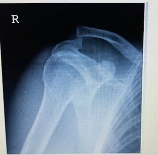
A 23-year-old man is brought to the emergency department 3 hours after injuring his shoulder playing basketball with his friends. Physical examination shows a step-off at the shoulder on palpation. An x-ray shows a total separation of the shoulder (see image below). Which of the following structures has most likely been torn?
A 22-year-old G1P0, at 32 weeks of gestation was admitted urgently to the hospital in preterm labor. Physical examination shows a breech footling presentation. The baby is delivered vaginally with considerable amounts of traction. The newborn is shown in image below. Which of the following structures was most likely injured during the childbirth?

A 56-year-old woman is brought to the emergency department after being involved in a motor vehicle collision. A large area of her anterior chest wall needed to be surgically removed and replaced with a musculo-osseous scapular graft involving the medial border of the scapula. Which of the following arteries will most likely compensate for the blood supply to the entire scapula?
A 25-year-old man is brought to the emergency department because he fell on a slippery trail and injured his right upper limb. Physical examination shows abrasions over his triceps brachii muscle. Which of the following nerves innervates the triceps brachii muscle?
A 24-year old woman is brought to the emergency department after she slipped on wet pavement and fell against the curb, injuring her right arm. Physical examination shows loss of sensation of the posterior forearm and dorsal hand. An x-ray of the right upper limb shows a mid-body fracture of the humerus. Which pair of structures is most likely injured at the fracture site?
A 20 year old man is brought to the emergency department after a motorbike collision. He is awake, alert, and oriented and his vital signs are within normal limits. Physical examination shows several cuts and bruises on his body. He is unable to extend the left wrist. Extension of the elbow is normal bilaterally. Sensation is lost in the lateral half of the dorsum of the left hand. Which of the following nerves is most likely injured, and in what part of the arm is the injury located?
A 27-year-old man is brought to the emergency department because of pain and swelling of his right arm after falling from a ladder. He is unable to abduct his right arm more than 15 degrees and resists lateral rotation due to pain. The patient also has a loss of sensation over the right shoulder. An x-ray of the arm shows an oblique fracture of the humerus. The most likely cause of these symptoms is a fracture affecting which of the following locations?
A 23-year-old man comes to the physician because of pain in the right upper extremity for several weeks. The pain is sharp, exacerbated by movement, and is progressively worsening since he increased his training for the upcoming basketball season. Examination of the right upper extremity shows no deformity. The pain is reproduced during the initial phase of abduction of the right arm, and when the arm is abducted against resistance. There is weakness in lateral rotation of the right shoulder. Which nerve was most likely injured?
A 44-year-old man comes to the physician after sustaining a penetrating wound to his shoulder from a crossbow bolt. Physical examination shows a deep, 4 cm laceration of the anterior shoulder and asymmetry of the radial artery. A compress is placed on the wound and deep pressure is applied. An angiogram shows transection of the axillary artery just distal to the origin of the subscapular artery. What collateral arterial pathways are available to bypass the site of injury?
A 45-year-old woman comes to the emergency department with a 2-week history of neck pain radiating to the left shoulder. Physical examination shows weakness in wrist extension and paresthesia on the back of her arm and forearm. An MRI examination finds a herniated disc in the cervical region. Which of the following spinal nerves is most likely injured?
A 60-year old woman comes to the physician because of intermittent numbness and tingling sensation of her right hand, which often wakes her up at night. Physical examination shows flexion of the wrist for 60 seconds reproduces the painful symptoms. A 3-month trial of wrist splinting and non-steroidal anti-inflammatory drugs therapy did not relieve the pain. During surgery, an anesthetic injection into the axillary sheath is administered. The axillary sheath takes its origin from which of the following structures?
A 45-year-old woman is brought to the emergency department by her husband because of a 2-week history of progressively increasing neck pain. Her pulse is 80/min, respirations are 20/min, and blood pressure is 128/32 mm Hg. Physical examination shows pain with mobilization, restricted range of motion of the neck, and weakness on extension of the forearm at the elbow. An MRI examination finds a herniated disc in the cervical region. Which of the following spinal nerves is most likely compressed?
A 25-year-old woman is brought to the emergency department after being involved in a motor vehicle collision. Physical examination shows that the right arm appears swollen, pale, and cool. Radial pulse is absent and any movement of the arm causes severe pain. An x-ray of the right arm shows a fracture at the radial groove of the humerus and a cast is placed. Three days later she has severe pain over the length of her arm. Which of the following conditions will most likely explain the findings on physical examination?
An 18-year-old man is brought to the emergency department because of left arm pain after an injury while playing rugby. Physical examination shows significant bruising over the left arm. An x-ray of the arm shows a transverse fracture of the humerus about 1 inch proximal to the epicondyles. Which of the following nerves is most likely injured by the jagged edges of the broken bone at this location?
A 17 year old girl is brought to the emergency department after sustaining a knife wound to her left arm in a street fight. Physical examination shows a 4 cm wound on the proximal medial arm. She has weakness of elbow flexion and supination of the left hand. Extension at the elbow and wrist is normal. Which of the following additional findings would be present on physical examination?
A 52 year old man comes to the emergency department with excruciating pain in the posterior aspect of his right forearm. For the past several days he has been rehearsing with the symphony orchestra as a conductor. Physical examination shows excruciating pain upon palpation 2 cm distal and posteromedial to the lateral epicondyle. Intramuscular steroids are administered. Which of the following is the most likely mechanism of injury?
A 58-year-old man is brought to the emergency department after an attempted robbery in which he sustained a bullet wound to the medial side of the elbow. His vital signs are within normal limits. A major nerve was repaired at the site where it passed behind the medial epicondyle. Bleeding was stopped from an artery which plays an important role in supplying blood supply to the nerve. Which of the following arteries was most likely repaired?
A 32-year-old woman comes to the hospital for hemodialysis. Physical examination shows venous access in her upper limb was unexpectedly difficult as the caliber of the major vein on the lateral aspect her arm was too small. A vein was found on the medial side of the arm that passed through the superficial and deep fascia to join veins beside the brachial artery. Which of the following veins is most likely located?
A 74-year-old woman is brought to the emergency department because of pain and swelling of her forearm after stumbling and falling over her pet dog. Physical examination of the forearm shows no open wounds and the neurovascular examination is within normal limits. The patient holds her left forearm in the pronated position and is unable to supinate the left hand. An x-ray of the right forearm shows a fracture of the upper third of the radius. The proximal end of the fracture deviates laterally. Which of the following muscles is primarily responsible for the lateral deviation?
A 43-year-old woman comes to the physician because of a 1-week history of elbow pain. She has pain over the lateral aspect of the right elbow that radiates to the posterior forearm. Physical examination shows tenderness distal to the origin of the extensor carpi radialis brevis. A diagnosis of lateral epicondylitis is made. Which of the following tests should be performed during physical examination to confirm the diagnosis?
A 54-year-old woman was brought to the emergency department after being found unconscious at the scene of a motor vehicle collision. Spine injury was suspected, and her neck was immobilized in a cervical collar. On arrival, her temperature is 37°C (98.6°F), pulse is 120/min, respirations are 16/min, and blood pressure is 142/84 mm Hg. Physical examination shows unilateral absence of her brachioradialis reflex. Injury to which of the following spinal nerves is most likely responsible for the absence of this reflex?
A 28-year-old woman is brought to the emergency department after experiencing severe pain in her chest while lifting weights. The pain was substernal and radiated to the mandible and her left arm. The woman felt dizzy and after 10 minutes she collapsed and became unconscious. A physician attempted to locate her radial pulse. The radial artery lies between two tendons near the wrists. Which of the following is the correct pair of tendons?
A 3-year-old girl is brought to the emergency department because of severe pain in her left upper extremity. Her mother recalls that she violently lifted the girl by her raised arm to prevent her from walking in front of a moving car. Physical examination shows that the patient’s forearm is pronated and extended at the elbow and she resists any movement due to pain. Which of the following is the most likely cause of the child’s pain?
A 41-year-old woman is brought to the emergency department after being involved in a motor vehicle collision. Vital signs are within normal limits. Physical examination shows a laterally deviated proximal portion of the radius. An x-ray of the forearm shows a transverse fracture of the radius proximal to the attachment of the pronator teres muscle. Which of the following muscles is most likely responsible for this deviation?
A 33-year-old man is brought to the emergency department with severe traumatic injuries after being involved in a motor vehicle collision. His temperature is 37°C (98.6°F), pulse is 115/min, and blood pressure is 89/39 mm Hg. A central venous line is placed for fluid resuscitation. Which of the following injuries is most likely to occur if a subclavian central venous line procedure is incorrectly performed?