Loading...
M1 Mediastinum Pre Formative Quiz
Quiz by Jenny Ousley
Tag the questions with any skills you have. Your dashboard will track each student's mastery of each skill.
A 62 year old man comes to the physician because of a 3 month history of progressive voice softening (hoarseness). Vital signs are within normal limits. Physical exam shows clear lung sounds bilaterally and no murmurs are heard. A CT scan of the chest shows a growth located within the arch of the aorta adjacent to the left pulmonary artery. Which neural structure is most likely being compressed to cause the changes in the patient’s voice?
A 42-year-old woman is brought to the emergency department by her husband because of 2-month history of hoarseness. Vital signs are within normal limits. Physical examination shows normal lung sounds bilaterally. A CT scan of the chest shows a mass at the aorticopulmonary window. Which of the following nerves is most likely compressed?
A 32-year-old woman is brought to the emergency department because of 5-hour history of severe dyspnea and anxiety. For the past 2 months she has been on a liquid diet because of dysphagia and has lost 15 kg (33 lb). Over the past several weeks, she has had bloody sputum during attacks of coughing and hoarseness. Fluoroscopy and a barium swallow show a 4-cm mass in the trachea compressing the thoracic esophagus. Which of the following nerves is most likely to be affected?
A 35-year-old man comes to the physician because of a 4-month history of progressive difficulty with swallowing solid food. Vital signs are within normal limits. Physical examination shows a diastolic murmur at the apex of the heart. A CT scan of the chest shows a dilated left atrium. Which structure is most likely being compressed by the expansion of the left atrium to result in the patient’s symptoms?
A 69-year-old woman is admitted to the hospital because of laryngeal cancer. Physical examination shows a thin-appearing woman with a hoarse voice. A CT scan examination of the chest and abdomen shows multiple masses in the lungs and liver. For nutritional needs a nasogastric tube is inserted. What is the last site at which resistance would be expected as the tube passes from the nose to the stomach?
A 47 year old man is brought to the emergency department because of difficulty swallowing. Physical exam shows swelling of the lower extremities. A barium swallow shows esophageal dilation with severe inflammation due to constriction at the esophageal hiatus from a large lipoma. Which of the following is the most likely cause of the severe edema of the lower limbs?
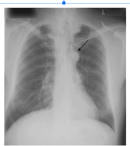
A 41-year-old man is brought to the emergency department with complaints of shortness of breath, dizziness, and sharp chest pain. Patient has a long history of smoking and hypertension. His temperature is 37°C (96.8°F), pulse is 120/min, respirations are 24/min, and blood pressure is 100/65 mm Hg. The large arrow in the x-ray of the chest indicates the region of pathology (Fig. 2.1). What is this structure?