Loading...


Cell Bio Week 10
Quiz by Haniya Hopson
Tag the questions with any skills you have. Your dashboard will track each student's mastery of each skill.
Actin filaments are said to “push” on the cell membrane to cause it to form a protrusion (see the figure). But there are problems with a pushing mechanism at both ends of the filaments. How can new subunits be added to the filament if the plus end of the filament must contact the membrane to push on it? And how is the minus end of the filament anchored so that the filament isn’t simply pushed back into the cell’s interior?How do you suppose new subunits are added to the filament to allow the plus end to push on the plasma membrane?
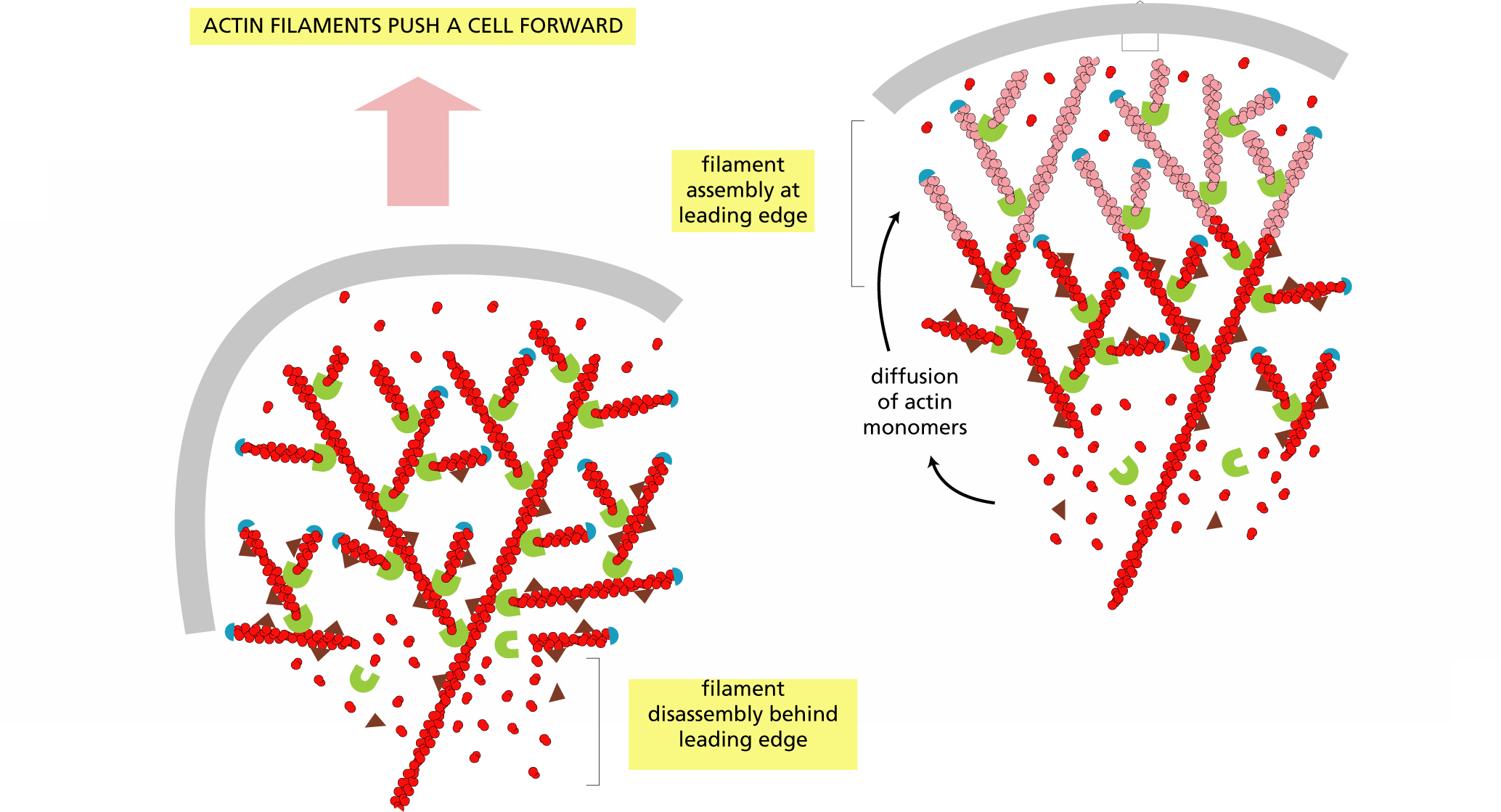
New subunits are added to plus ends exposed by thermal motion.
New subunits are added into an interior location in the filament.
New subunits are added to branches made by the Arp2/3 complex.
New subunits are added to the opposite (minus end) of a filament.
A typical time course of polymerization of actin filaments from actin subunits is shown in the figure. This experiment used purified actin monomers (with bound ATP) that were labeled with a fluorescent probe. Upon polymerization, the fluorescence of the probe increases, which allows actin polymerization to be measured.If you doubled the initial concentration of actin, how do you suppose the concentration of free actin at equilibrium would change relative to the original experiment?

Free actin concentration would be the same as in the original experiment.
Free actin concentration would be higher than in the original experiment.
Free actin concentration would be lower than in the original experiment.
Free actin concentration could be more or less than in the first experiment.
Actin filaments are said to “push” on the cell membrane to cause it to form a protrusion (see the figure). But there are problems with a pushing mechanism at both ends of the filaments. How can new subunits be added to the filament if the plus end of the filament must contact the membrane to push on it? And how is the minus end of the filament anchored so that the filament isn’t simply pushed back into the cell’s interior?How do you suppose new subunits are added to the filament to allow the plus end to push on the plasma membrane?
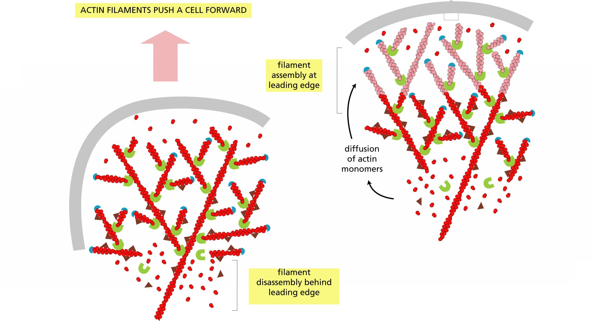
A typical time course of polymerization of actin filaments from actin subunits is shown in the figure. This experiment used purified actin monomers (with bound ATP) that were labeled with a fluorescent probe. Upon polymerization, the fluorescence of the probe increases, which allows actin polymerization to be measured.If you doubled the initial concentration of actin, how do you suppose the concentration of free actin at equilibrium would change relative to the original experiment?

What is the term for the process by which a polymeric protein filament gains subunits at one end while simultaneously losing subunits from the other end?
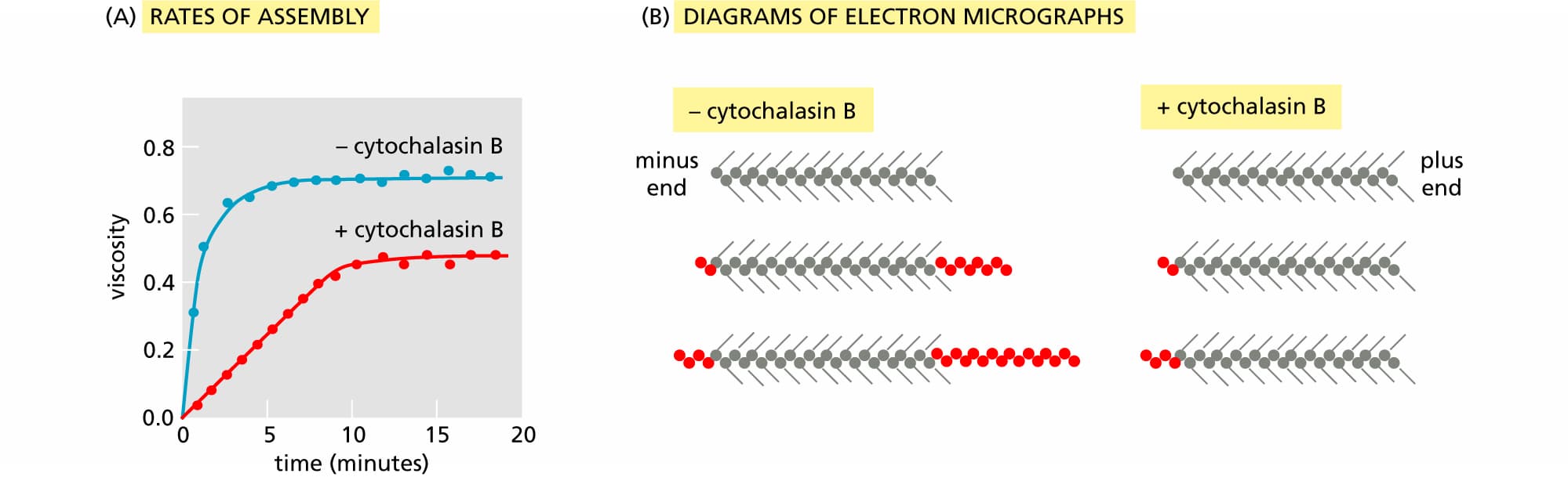
Cytochalasin B interferes with the assembly of actin filaments. In the classic experiment that defined its mechanism, short lengths of actin filaments were decorated with myosin heads and then mixed with actin subunits in the presence or absence of cytochalasin B. Assembly of actin filaments was measured by assaying the viscosity of the solution (see the figure Part A) and by examining samples by electron microscopy (Part B). The decorated actin filaments present before the addition of actin monomers are shown at the top of each set of three. Filaments present after increasing times of incubation with actin monomers (red circles) are shown below.Which of the following statements describes a plausible mechanism of cytochalasin B action that accounts for the slower rate of assembly and the appearance of the filaments in the electron micrographs?
Some actin-binding accessory proteins significantly increase the rate at which the formation of actin filaments is initiated in the cytosol. How might such proteins do this? What must they not do when binding the actin monomers?
What is the term for the protein assembly that nucleates actin filament growth from the minus end, allowing rapid growth at the plus end and forming a treelike web of filaments?
Most eukaryotic cells maintain the concentration of actin monomers well above the critical concentration needed for actin polymerization in the test tube. In most cells, the excess actin monomers are bound by the protein profilin, which regulates their addition to actin filaments.
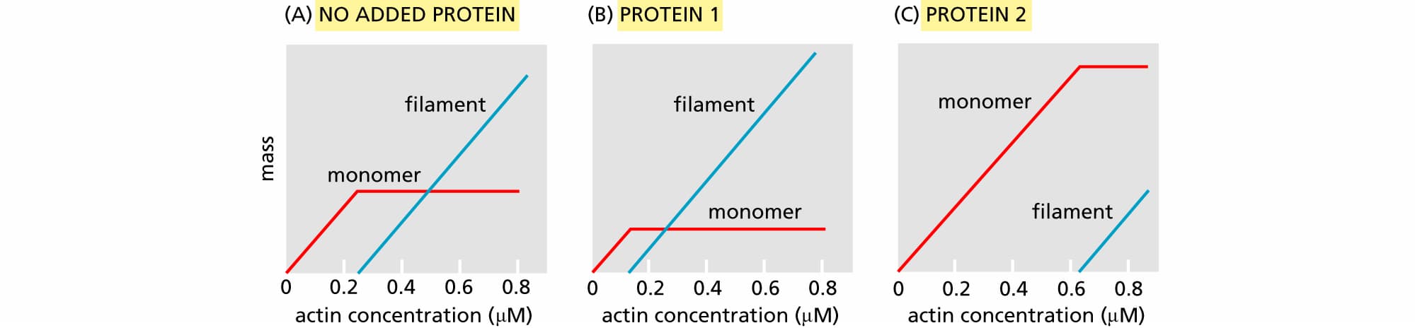
You have two proteins that you suspect cap the ends of actin filaments. To determine whether they do and, if so, which protein caps which end, you measure filament formation as a function of G-actin concentration (labeled 'actin concentration' on the x-axes) in the absence of either protein, in the presence of protein 1, and in the presence of protein 2 (see the figure). The mass of actin, as monomers or filaments, was determined at equilibrium.Which end of the actin filament is capped by protein 1? Which by protein 2?
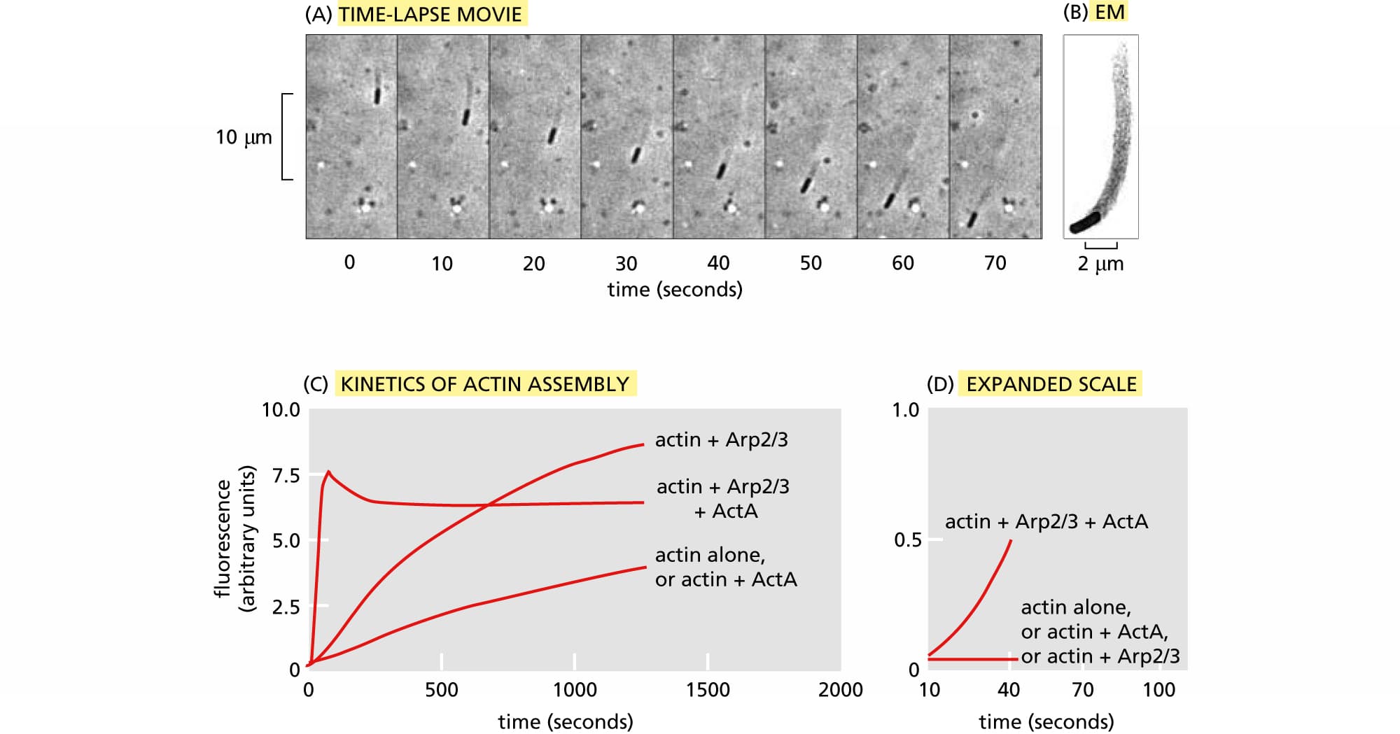
The intracellular pathogenic bacterium Listeria monocytogenes propels itself through the cytosol on a comet tail of actin filaments provided by the host cell, as shown by light microscopy (see the figure Part A) and electron microscopy (Part B). Remarkably, only a single bacterial protein, the transmembrane protein ActA, is required for this motility. ActA is distributed unequally on the surface of the bacterium, with maximum concentrations at the pole in contact with the actin tail.The effects of ActA on in vitro actin polymerization in the presence and absence of the Arp2/3 complex are shown in Part C, and the first few seconds of the reactions are shown on an expanded scale in Part D. Polymerization of actin was followed using a fluorescent tag that exhibits much higher fluorescence intensity when actin is polymerized.Which one of the following statements correctly describes the effects of ActA and the Arp2/3 complex, separately or together, on the rate of nucleation of actin filaments?
The adult animal seethes with crawling cells. Macrophages and neutrophils, for example, crawl to sites of infection and engulf foreign invaders as a critical part of the innate immune response.Which of the following statements correctly describes the individual processes that make up the crawling movement of cells?
I Protrusion pushes out the leading edge of the plasma membrane at the front end of the cell.
II Adhesion to the surface allows the internal actin cytoskeleton to connect to the substratum.
III Contraction of bundles of actin filaments and kinesin motor proteins at the rear of the cell enables translocation.
Myosin II bipolar thick filaments slide actin filaments toward one another to bring about muscle contraction. What arrangement of actin filaments is necessary for myosin to bring about contraction?
Match each of the following events with the correct binding to or release of nucleotide from myosin.
Rigor mortis occurs in muscle cells after death and is characterized by muscles being locked in a contracted state where myosin is bound to actin. What could be added to the muscle cell to release the rigor state?
Which one of the following changes takes place when a skeletal muscle contracts?
Place the following steps of muscle contraction in the order in which they occur.
Which of the following would increase the level of muscle contraction?
Except for one single type of myosin motor protein, all other members of the 37 distinct families in the myosin superfamily move toward the plus end of an actin filament.
Which of the following statements are consistent with the structure and function of intermediate filaments?
Mutation of the muscle-specific intermediate filament desmin leads to the rare disease desmin-related myopathy. This disorder starts with weakness of the lower limbs when patients are in their 20s or 30s. As symptoms worsen, weakness in respiratory and cardiac muscles occurs, which can lead to serious problems including sudden cardiac arrest.Which of the following mutations would disrupt desmin intermediate filament structure or function and could explain the symptoms of desmin-related myopathy? Choose all of the possible mutations.
There are no known motor proteins that move on intermediate filaments. Which one of the following hypotheses best explains this observation?